Few biological facts seem as irrevocable as brain death. It has long been assumed that when we die, our neurons die with us. But a new study on the neuron-packed tissue of the eye is beginning to challenge that dogma.
In the new work, researchers restored electrical activity in human retinas—the light-sensitive neural tissue that sits at the back of our eyes and communicates with our brains—from recently deceased organ donors. This achievement, reported in Nature, offers a better way to study eye diseases such as age-related macular degeneration, a leading cause of vision loss and blindness. It could also lay the groundwork for reviving other types of neural tissue and perhaps—one day—for retinal transplants.
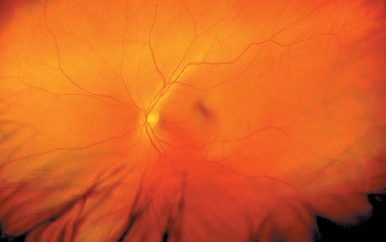
Most retina studies are done in animals, primarily mice. But mouse retinas lack the macula, a key region found in human eyes that picks out fine details, so they are not an ideal model. Human eye tissue from autopsies often takes hours to obtain and is dead before scientists can study its function. But what if you could revive it?
When Yale University researchers showed in 2019 that rudimentary electrical activity could be restored in pig brains after death, University of Utah vision scientist Frans Vinberg, Scripps Research retinal surgeon Anne Hanneken and their colleagues were inspired to study whether retinal tissue could also be restored postmortem.
For the study, the researchers first tested how long mouse retinas could send electrical signals after the animals were euthanized. They were able to restore this activity up to three hours later—and found that a lack of oxygen was the main factor in irreversible loss of function. They then investigated human eyes that the researchers obtained from organ donors very soon after brain or cardiac death. The scientists transported the eyes to the laboratory in a container that supplied oxygen and nutrients, then exposed the retinal tissue to dim light and measured electrical signals generated by the tissue. They were able to reestablish electrical activity in light-sensitive cells called photoreceptors, as well as in the neurons these cells connect to, in the donor eyes—if the eyes were obtained less than 20 minutes after death. Of course, the eyes could not “see,” because they were not connected to a brain, Hanneken notes. But the results showed it was possible to restore not just individual retinal cells but the communication between them.
“What’s most exciting is this really could become a model for studying visual physiology in human retinas, in health and in aging and in disease,” says Joan Miller, chief of ophthalmology at Mass Eye and Ear and ophthalmology chair at Harvard Medical School, who was not involved with the new study. Macular degeneration, for example, has so far been difficult to study because living human eye tissue has been impossible to access. Using this new technique, scientists could study healthy and diseased donor eyes to understand their function and test treatments.
The team’s findings also suggest it may be possible to revive other types of neural tissue. “The retina is a window to the brain, so if you can restore communication in the retina after death, it makes you pause and consider what kind of communication you might be able to recover in the brain,” Hanneken says. The study additionally raises the prospect of retinal transplants, although those are likely still very far off, the researchers say.
This new work illustrates the importance of donor tissue to basic science. “We are very thankful for the donors and their families,” Vinberg says. “We hope this will encourage people … to check that box in their driver’s license and also be willing to donate tissues for research.”